


SEEFOR 17(2): 26019
Article ID: 26019
DOI: https://doi.org/10.15177/seefor.26-019
ORIGINAL SCIENTIFIC PAPER
Influence of Individual Tree Variability and Branch Age of Wild Cherry (Prunus avium) on Damage Caused by Diaporthe eres
Aleksandar Vemić1, Sanja Lazić2, Ljubinko Rakonjac3, Aleksandar Lučić2, Snežana Stajić3, Marina Vukin4, Vladan Popović2*
Addresses:
(1) Institute of Forestry, Department of Forest Protection, Kneza Višeslava 3, RS-11000 Belgrade, Serbia;
(2) Institute of Forestry, Department of Genetics, Plant Breeding, Seed and Nursery Production, Kneza Višeslava 3, RS-11000 Belgrade, Serbia;
(3) Institute of Forestry, Department of Forest Establishment, Silviculture and Ecology, Kneza Višeslava 3, RS-11000 Belgrade, Serbia;
(4) University Metropolitan, Faculty of Applied Ecology "Futura", Požeška 83a, RS-11000 Belgrade, Serbia
Citation: Vemić A, Lazić S, Rakonjac Lj, Lučić A, Stajić S, Vukin M, Popović V, 2026. Influence of Individual Tree Variability and Branch Age of Wild Cherry (Prunus avium) on Damage Caused by Diaporthe eres. South-east Eur for 17(2): 26019. https://doi.org/10.15177/seefor.26-019.
Received: 3 May 2026; Revised: 14 Jul 2026; Accepted: 16 Jul 2026; Published online: 27 Jul 2026
Cited by: Google Scholar
Abstract
The occurrence of canker on wild cherry (Prunus avium) caused by Diaporthe eres raises several unresolved questions regarding the pathogen's pathogenicity, symptom development, disease progression, and management. This study demonstrated that symptom development differed among branch age classes and individual host trees. Dieback occurrence and necrotic lesion dimensions were analysed at the end of the trial, after 81 days. Perennial branches had the largest dimensions of necrotic lesions, followed by annual branches. The smallest necroses were recorded on leading shoots. There was a statistically significant difference in the dimensions of necrotic lesions and branch dieback at the individual tree level. Tolerant individual trees showed fewer necrotic lesions and lower dieback than sensitive individual trees. This study showed, for the first time, that Diaporthe eres symptoms develop differently on wild cherry depending on the part of the tree and the individual's tolerance to this pathogen. The results can be applied to improve monitoring strategies for Diaporthe eres and support further work on selecting and producing wild cherry trees tolerant to Diaporthe eres.
Keywords: susceptibility; selection; canker suppression; protection
INTRODUCTION
Wild cherry (Prunus avium (L.) L.) is a deciduous, medium-sized, short-lived, fast-growing species of exceptional decorative value. It is used as food and as a raw material for the production of parquet, furniture, and musical instruments (Welk et al. 2016). Various organs of wild cherry trees are rich in bioactive components used in the food and pharmaceutical industries (Jesus et al. 2020, Hu et al. 2021, Willig et al. 2022, Clodoveo et al. 2023).
Global changes influence plant pathogens, as evidenced by changes in forest health dynamics (Pautasso et al. 2015). Many unidentified fungal species have been recorded on wild cherry trees (Bien and Damm 2020). Additionally, new research indicates that the Diaporthe eres species complex causes cankers on wild cherry trees in China (Chen et al. 2023, Dai et al. 2024).
Fungi of the genus Diaporthe are parasites, saprophytes, or endophytes, and are dispersed worldwide (Udayanga et al. 2014, Dissanayake et al. 2017). In forestry, the most common hosts for pathogens from the genus Diaporthe belong to the genera Acer, Eucalyptus, Fraxinus, Juglans, Prunus, Robinia, Rosa, Sambucus, Sorbus, Ulmus, etc. (Santos et al. 2017, Yang et al. 2018, Abeywickrama et al. 2022, Zabiák et al. 2023a).
The mechanisms of infection caused by species of the Diaporthe genus depend on the host's defensive reactions, the degradation degree and modification of the host's cell wall, as well as detoxification of compounds, transporter activities, and toxin production (Mena et al. 2022). The Diaporthe eres species complex produces many metabolites, including phytotoxic compounds such as cyclopaldic acid, altiloxin A and B, and their derivatives (Abramczyk et al. 2023). It also produces enzymes, the most common of which are hydrolases and oxidoreductases (Hilário et al. 2022).
Further studies of the Diaporthe eres pathogenesis in wild cherry are necessary to prevent and reduce damage to trees that may occur due to its spread. Firstly, it is necessary to determine the role of individual trees and branch age in the development of necrotic lesions (i.e., cankers). In woody plants, young branches exhibit higher rates of respiration and cell division, as well as higher concentrations of growth hormones and a more active phloem than older branches (Evert 2006). Moreover, young branches contain higher levels of phenolic compounds, which play a key role in plant defense responses (Feucht et al. 1986, Evert 2006). In contrast, older branches contain a greater proportion of lignified fibers, thicker cell walls, and fewer living parenchyma cells than younger branches, and their bark is thicker, providing greater mechanical resistance (Evert 2006). Thus, young tissues rely primarily on stronger chemical defenses, whereas structural defenses are more pronounced in older tissues. Given these physiological and anatomical differences among leading shoots, annual branches, and perennial branches, it is important to investigate whether branch age influences susceptibility to the poorly studied pathogen Diaporthe eres in wild cherry. Understanding this relationship will provide insight into the decline of wild cherry trees and serve as a foundation for developing protection methods that do not rely on chemical treatments. Also, the findings can represent a starting point for further research of different cultivars and varieties of wild cherry, as well as cultivation measures that promote the growth of specific branch types.
In light of the above rationale, this study examined the pathogenicity of Diaporthe eres on wild cherry branches of various ages. The research was conducted in the living archive of the Lipovica Research Station. The tested null hypotheses were as follows: (i) there is no difference in the susceptibility of wild cherry individual trees to Diaporthe eres; (ii) there is no difference in the dimensions of necrotic lesions caused by Diaporthe eres between leading shoots, annual, and perennial branches of wild cherry.
MATERIALS AND METHODS
Plant Material
The study included ten naturally regenerated wild cherry (Prunus avium) trees growing in a natural stand of Hungarian oak (Quercus frainetto Ten.) and Turkey oak (Quercus cerris L.). Test trees were located at the "Lipovica" research station of the Institute of Forestry in Belgrade, Serbia (44°38'06''N, 20°24'50''E, 285 m.a.s.l.). The trees originated from the same local population and grew under similar ecological conditions. Trees of approximately the same age, size, and health status were selected to minimize the influence of ontogenetic and physiological differences on disease development. The trees were estimated to be approximately 20 years old. For three representative trees, age was determined by counting annual growth rings in increment cores collected with an increment borer, whereas the age of the remaining trees was estimated based on their similar dimensions, growth habits, and the approximate timing of their natural regeneration. The wild cherry trees were isolated trees, thereby reducing the risk of natural infection by other pathogens. Tree health status was assessed in accordance with the ICP Forests Manual (2020–2022), the version in force at the time of the experiment. The selected trees appeared visually healthy. They showed no signs of damage.
Wild Cherry Inoculation
The experiment was conducted from June to September 2024. The mean air temperature during the experimental period was 25.4°C, the mean relative humidity was 1.7% (RHMSS 2025), and the cumulative precipitation was 204.7 mm (RHMSS 2025). A total of 186 branches were inoculated, with 10 to 20 branches from each wild cherry tree. This included 49 leading shoots, 48 annual branches, and 89 perennial branches. A smaller number of leading shoots and annual branches than perennial branches were inoculated in order to preserve the young parts of the crown and prevent crown deformation following completion of the experiment. The Diaporthe eres isolate (strain U11MNE, GenBank accession numbers MK352454 and MK358120) from the mycological collection of the Institute of Forestry in Belgrade was used for this trial. The isolate was selected for its pronounced pathogenicity (Vemić 2020) and to reduce variability within the tested material (Udayanga et al. 2014). This increased the precision of data analysis. Cultures of Diaporthe eres 10 days old, grown on 3% malt extract agar (MEA) nutrient medium (Biolab, Hungary; Torlak, Serbia), were used for inoculation. The inoculation method involved making a 5×5 mm bark incision approximately 2 mm deep in the middle of each branch with a sterile scalpel. Mycelium fragments measuring 5×5 mm were inserted into the incision. The inoculated places were wrapped with Parafilm® M laboratory film (Bemis Company Inc., Neenah, Wisconsin, USA) and aluminum foil. Additionally, a control group was established by inoculating 15 branches on each wild cherry tree with sterile, pure 3% MEA nutrient medium. This was done in the same manner as for Diaporthe eres inoculation. A total of 150 branches were inoculated in the control group.
The trial ended after 81 days. During this period, all inoculated trees showed disease symptoms. Inoculated branches were cut to the stem collar. The presence of dieback and the dimensions of necrotic lesions were analysed. The dieback on each branch was assessed visually. Before measuring the necrotic lesions, the bark was removed. Immediately after debarking, necrotic lesions were estimated based on cambium discoloration. The length of the necrotic lesions was measured vertically from one edge of discoloration to the other. The width was measured as the circumference at the points of the greatest discoloration. Branch mortality was estimated by observing the occurrence of wilted wood. The tested branches were classified as sensitive when they exhibited extensive necrotic lesion development, defoliation, and branch dieback, whereas branches were classified as tolerant when necrotic lesions remained localized and complete defoliation and branch dieback did not occur.
Re-isolation was tried in all branches used in the trial. The re-isolation of leading shoots and annual branches included cutting them into rings, coating them with 96% alcohol, and exposing them to a flame for a few seconds. After that, the bark was peeled off, and the procedure was repeated. These sections were placed on a 3% MEA nutrient medium. In the case of perennial branches, the parts with the appearance of necrotic lesions were cut into sections. Then, the part containing the necrotic lesion and the surrounding asymptomatic tissue was cut from each section. These fragments were sterilized like leading shoots and annual branches and mounted on a 3% MEA nutrient medium. In perennial branches from the control group, which did not develop necrotic lesions, randomly selected parts were sterilized and similarly placed on the nutrient medium. During re-isolation, the presence of Diaporthe eres was confirmed based on morphological characteristics, including colony appearance, growth pattern, sporulation, and conidial morphology, all of which were identical to those of the original isolate used for inoculation, which had previously been identified by molecular methods (Vemić 2020). The morphological characteristics of the re-isolates and the original isolate were compared according to the descriptions provided by Udayanga et al. (2014) and Vemić (2020).
Statistical Analysis
The dimensions of necrotic lesions within individual trees and branches of different ages were tested for normality of distribution using the Kolmogorov-Smirnov test with Lilliefors correction. In the same way, the normality of the residuals was tested for the application of the general linear model (GLM) and standard multiple regression. The homogeneity of variances was tested using Levene's test. Since the conditions for applying parametric tests and models were not met, non-parametric tests were used to compare the dimensions of necrotic lesions between different groups.
The Mann-Whitney U test was used to compare the dimensions of necrotic lesions between the Diaporthe eres inoculated group and the control group. The Kruskal-Wallis test was used to assess differences in the length and width of necrotic lesions between individual trees
inoculated with Diaporthe eres and the control group. Dunn's post hoc test was used to determine differences between different pairs of individual trees in necrotic lesion dimensions.
The Chi-Square Test of independence was used to test the association between branch inoculation with Diaporthe eres and mortality. The Z-test was used to compare dieback occurrence between individual trees and branches of different ages.
All statistical analyses were performed using Microsoft Excel 2021 and SPSS 27.
RESULTS
General Observations
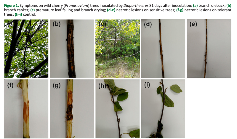
Wild cherry trees inoculated with Diaporthe eres exhibited dieback, cankers, and premature leaf shedding, often accompanied by branch mortality (Figure 1). The Mann-Whitney test showed that there was a statistically significant difference in the length of necrotic lesions (U = 3164,500, p˂0.001; Figure 1) and width of necrotic lesions (U = 8331,000, p˂0.001; Figure 1) between wild cherry branches inoculated with Diaporthe eres and branches in the control group. Re-isolation was successful from all branches inoculated with Diaporthe eres. In the control group, no re-isolation of Diaporthe eres was recorded.
Influence of Wild Cherry Individual Tree on Diaporthe eres Development
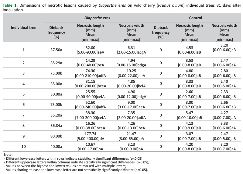
The Z-test showed a statistically significant difference in the proportion of branch dieback between the individual trees (Table 1, Figure 1). The Kruskal-Wallis test showed a statistically significant difference in the length (H = 40.306, p˂0.001) and the width of necrotic lesions (H = 48.127, p˂0.001) on the total number of branches between individual trees inoculated with Diaporthe eres (Table 1, Figure 1). Among individual trees, based on the total number of inoculated branches, 10% showed the shortest or longest necrotic lesions, while the rest were in the transition between the most tolerant and the most sensitive (Table 1). Therefore, the width of necrotic lesions showed similar variation, with 80% of the trees at the transition between the most sensitive and the most tolerant, as determined by the total number of inoculated branches (Table 1). That is, individual tree 9 was the most sensitive, while individual tree 10 was the least sensitive. In addition, 50% of the trees did not show a statistically significant difference in lesion width compared to the control group (Table 1).
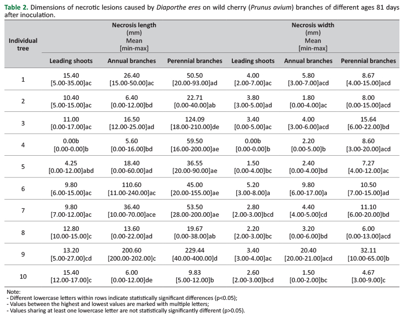
Moreover, between the individual trees, there was a statistically significant difference in the length of necrotic lesions and the width of necrotic lesions on leading shoots (H = 21.870, p˂0.001; H = 27.525, p˂0.001), annual branches (H = 29.674, p˂0.001; H = 34.804, p˂0.001) and perennial branches (H = 39.158, p˂0.001; H = 38.403, p˂0.001) based on the Kruskal-Wallis test (Table 2). However, only 10-20% of the individual trees showed the smallest length or width of necrotic lesions within a particular branch age, i.e., similar to the total number of inoculated branches (Table 1, Table 2).
Influence of Wild Cherry Branch Age on Diaporthe eres Development
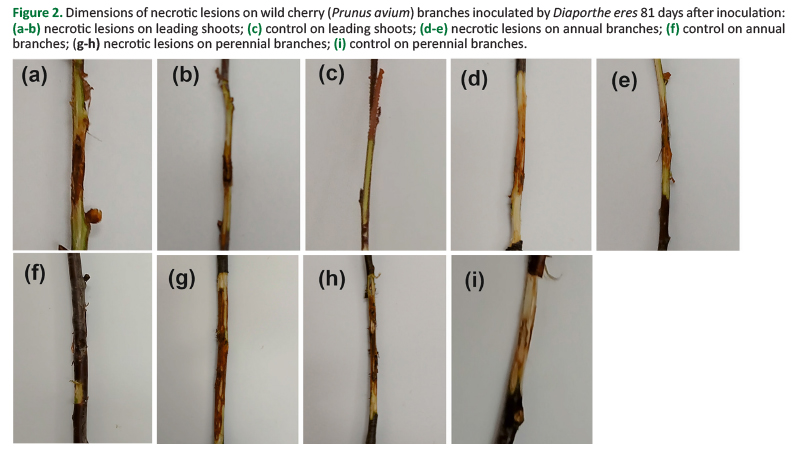
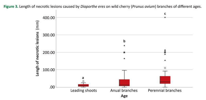
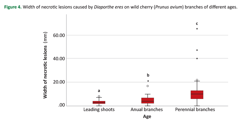
There was a statistically significant difference in the length (H = 60.445, p˂0.001; Figure 2; Figure 3) and width of necrotic lesions (H = 72.918, p˂0.001; Figure 2; Figure 4) between leading shoots, annual, and perennial branches of wild cherry.
The leading shoots had the smallest length and width of necrotic lesions, followed by annual branches, while perennial branches had the largest dimensions of necrotic lesions (Figure 2, Figure 3, Figure 4).
There was a statistically significant association between the inoculation of leading shoots, annual and perennial branches with Diaporthe eres and mortality (χ2 = 15.338, p˂0.001, Figure 1).
DISCUSSION
This study provides a more detailed examination of the influence of wild cherry individual trees and branch age on the development of Diaporthe eres. For the first time, it was found that cankers develop at different rates depending on the age of the branch on which they appear. Individual trees also showed differences in individual tolerance to the pathogen. Therefore, the first two null hypotheses were rejected. The alternative hypotheses were accepted: (i) there is a difference in the susceptibility of wild cherry individual trees to Diaporthe eres; (ii) there is a difference in the dimensions of necrotic lesions caused by Diaporthe eres between wild cherry leading shoots, annual, and perennial branches. This variability provides a valuable basis for future wild cherry selection and breeding programs.
One aspect that warrants attention, although it was not directly addressed in the present study, is the potential application of these findings to monitoring Diaporthe eres in wild cherry. Many models can be used to predict the spread of various pathogens affecting woody species (Oliva et al. 2013, Zhao et al. 2020, Webb et al. 2023). The prediction of Diaporthe eres development relies on understanding the influence of temperature, relative humidity, and precipitation (Thomidis and Michailides 2009, Arciuolo et al. 2021, Camardo Leggieri et al. 2022, Zabiák et al. 2023b). Therefore, in addition to these variables, models should also incorporate host tissue age and individual variability in wild cherry as predictive parameters. In any case, field monitoring can be directly improved. The interval between monitoring activities should be shorter for trees that have shown symptoms and have denser crowns composed of perennial branches. Additionally, monitoring can be further improved by prioritizing the inspection of perennial branches, i.e., the most susceptible parts of the crown, which enables earlier detection and reduces the number of inspections required within a stand or plantation. Future studies based on the proposed and related research directions will help determine the most effective approaches for predicting the spread and development of Diaporthe eres in wild cherry.
There is significant variability in the physiological, morphological, pomological, anatomical, and biochemical parameters of wild cherry (Khadivi et al. 2019, Miljković et al. 2019, Azizi-Gannouni et al. 2020, Corneanu et al. 2020, Stojnić et al. 2022, Dangi et al. 2024). Specifically, genetic variability exists among different trees of this species (Campoy et al. 2016, Patzak et al. 2019). Therefore, it is necessary to utilize the existing gene pool by selecting trees that naturally confer tolerance within local populations. A reduction in genetic diversity has been observed in isolated stands (Lobo et al. 2018). In such stands, the introduction of wild cherry individual trees tolerant of Diaporthe eres may be justified. However, source and reproductive material should be selected from ecologically similar populations to avoid adverse effects on the genetic pool of the trees. The necrosis width in half of the individual trees did not differ from that of the control group, whereas the lesion length in all individual trees was greater than that of mechanical-damage-induced lesions in the control group. Therefore, both parameters should be considered during selection to avoid errors in assessing suitable individual trees. Since this study showed that necrosis intensity varied with branch age, we believe that the technique and intensity of pruning can be of great importance in protecting wild cherry trees from the development of cankers caused by Diaporthe eres. This may be beneficial given that selective pruning of wild cherry does not negatively affect tree growth (Sprengel et al. 2018, Fernández-Moya and Urbán-Martínez 2022).
Wild cherry fruits can be extensively damaged by Diaporthe eres (Liu et al. 2024). Additionally, various fungi, viruses, and bacteria damage the trees and fruits of wild cherry (Marković 2012, Serradilla et al. 2021, Reinhold and Pscheidt 2023, Cancino et al. 2023, Marroni et al. 2024, Liu et al. 2024). Furthermore, certain species from the genus Phytophthora cause root rot and infection symptoms in the wild cherry crown (Kurbetli 2014, Milenković 2015, Jung et al. 2016). Some of the fungicides effective in controlling Diaporthe eres are prochloraz, tebuconazole, pyraclostrobin, thiophanate-methyl, carbendazim, and thiram (Thomidis and Michailides 2009, Król et al. 2019, Tao et al. 2020). Some other substances, such as silver nitrate and AgSe nanoparticles, have also been used to control Diaporthe eres (Štůsková et al. 2022). We believe it is necessary to investigate whether measures based on individual tree selection and the regulation of the number of branches of different ages can reduce symptoms caused by other agents associated with wild cherry decline and reduce fungicide use.
In summary, the results of this study indicate that although differences among individual trees were observed and, in some cases, substantial, they were limited compared to those among branches of different ages. Additionally, a limitation of this study is that it relies on a single isolate of Diaporthe eres, which allows for assessing the effects of individual tree and branch age on host susceptibility; however, to investigate the response of wild cherry populations, it is necessary to include multiple strains of Diaporthe eres. Accordingly, management strategies should not rely solely on either of these factors. As indicated above, a combined approach that includes selecting more tolerant individuals and regulating crown structure may improve monitoring efficiency and protect wild cherry from Diaporthe eres, while potentially enhancing control measures against other diseases affecting wild cherry.
CONCLUSIONS
This study examined wild cherry trees' tolerance to the development of cankers caused by Diaporthe eres. The results indicated differences in the size (length and width) of necrotic lesions caused by this pathogen across all branches and among branches of varying ages. Furthermore, leading shoots, annual branches, and perennial branches of wild cherry developed cankers, which varied in size across individual trees. Most wild cherry individual trees showed similar tolerance to Diaporthe eres. Therefore, the developmental stage of the tissue may be more important for the development of Diaporthe eres than host origin. The larger cankers were recorded on perennial branches, with the size decreasing as the age of the branches decreased. The findings enable a better understanding of the Diaporthe eres bioecology and, as such, have wide application in further studies of this pathogen and in reducing damage to wild cherry trees.
Author Contributions
AV, VP conceived the research, AV, VP and SL designed the research, AV and SL carried out the field measurements, AV and SL performed laboratory analysis, AV, SL, LjR, AL, SS, MV processed the data and performed the statistical analysis, LjR, AL secured the research funding, supervised the research and helped to draft the manuscript, AV, VP and SL wrote the manuscript.
Funding
This research was funded by the Ministry of Science, Technological Development and Innovation. Contract No. 451-03-33/2026-03/200027.
Conflicts of Interest
The authors declare no conflict of interest.
REFERENCES
Abeywickrama PD, Camporesi E, Jayawardena RS, Hyde KD, Yan J, Zhang W, Li X, 2022. Novel and Surprising Host Associations of Diaporthe (Diaporthaceae, Diaporthales) species from Italy. Chiang Mai J Sci 49(2): 223-247. https://doi.org/10.12982/CMJS.2022.028.
Abramczyk B, Pecio Ł, Kozachok S, Kowalczyk M, Marzec-Grządziel A, Król E, Gałązka A, Oleszek W, 2023. Pioneering Metabolomic Studies on Diaporthe eres Species Complex from Fruit Trees in the South-Eastern Poland. Molecules 28(3): 1175. https://doi.org/10.3390/molecules28031175.
Arciuolo R, Camardo Leggieri M, Chiusa G, Castello G, Genova G, Spigolon N, Battilani P, 2021. Ecology of Diaporthe eres, the causal agent of hazelnut defects. PLOS ONE 16(3): e0247563. https://doi.org/10.1371/journal.pone.0247563.
Azizi-Gannouni T, Sghaier T, Ammari Y, 2020. Behavior and morphometric characterization of local and introduced cultivars of sweet cherries (Prunus avium), tested in a multi-site trial in Tunisia. Sci Hortic 270: 109455. https://doi.org/10.1016/j.scienta.2020.109455.
Bien S, Damm U, 2020. Prunus trees in Germany—a hideout of unknown fungi? Mycol Prog 19(7): 667-690. https://doi.org/10.1007/s11557-020-01586-4.
Camardo Leggieri M, Arciuolo R, Chiusa G, Castello G, Spigolon N, Battilani P, 2022. DEFHAZ: A Mechanistic Weather-Driven Predictive Model for Diaporthe eres Infection and Defective Hazelnut Outbreaks. Plants 11(24): 3553. https://doi.org/10.3390/plants11243553.
Campoy JA, Lerigoleur-Balsemin E, Christmann H, Beauvieux R, Girollet N, Quero-García J, Dirlewanger E, Barreneche T, 2016. Genetic diversity, linkage disequilibrium, population structure and construction of a core collection of Prunus avium L. landraces and bred cultivars. BMC Plant Biol 16: 49. https://doi.org/10.1186/s12870-016-0712-9.
Cancino S, Lolas M, Galdós L, Hernández Y, Ferrada E, Riveros P, Blanco-Ulate B, Díaz GA, 2023. Occurrence of Alternaria alternata and A. tenuissima Causing Black Rot in Cherry Fruits (Prunus avium) in Central Chile. Plant Dis 107(12): 4032. https://doi.org/10.1094/PDIS-07-23-1435-PDN.
Corneanu M, Iurea E, Sîrbu S, 2020. Biological properties and fruit quality of sweet cherry (Prunus avium L.) cultivars from Romanian assortment. Agron Res 18(4): 2353-2364. https://doi.org/10.15159/AR.20.231.
Chen P, Abeywickrama PD, Ji S, Zhou Y, Li X, Zhang W, Yan J, 2023. Molecular Identification and Pathogenicity of Diaporthe eres and D. hongkongensis (Diaporthales, Ascomycota) Associated with Cherry Trunk Diseases in China. Microorganisms 11(10): 2400. https://doi.org/10.3390/microorganisms11102400.
Clodoveo ML, Crupi P, Muraglia M, Naeem MY, Tardugno R, Limongelli F, Corbo F, 2023. The main phenolic compounds responsible for the antioxidant capacity of sweet cherry (Prunus avium L.) pulp. LWT 185: 115085. https://doi.org/10.1016/j.lwt.2023.115085.
Dai Q, Zhang Q, He M, Ai J, Cai F, 2024. Occurrence and molecular characterization of Diaporthe eres causing stem canker on sweet cherry trees (Prunus avium) in northern China. Plant Pathol 73(7): 1794-1800. https://doi.org/10.1111/ppa.13929.
Dangi G, Singh D, Chauhan, N, Dogra RK, Verma P, Chauhan A, 2024. Evaluating genetic diversity of morpho-physiological traits in sweet cherry (Prunus avium L.) cultivars using multivariate analysis. Genet Resour Crop Evol 71: 3267-3302. https://doi.org/10.1007/s10722-023-01809-z.
Dissanayake AJ, Phillips AJL, Hyde KD, Yan JY, Li XH, 2017. The current status of species in Diaporthe. Mycosphere 8(5): 1106-1156. https://doi.org/10.5943/mycosphere/8/5/5.
Evert RF, 2006. Esau’s Plant Anatomy: Meristems, Cells, and Tissues of the Plant Body: Their Structure, Function, and Development. 3rd Edition. John Wiley & Sons, Hoboken, New Jersey, 601 p. https://doi.org/10.1002/0470047380.
Fernández-Moya J, Urbán-Martínez I, 2022. Growth performance of wild cherry (Prunus avium L.) clones in planted forests under different managements in Galicia, NW Spain. Silvae Genet 71(1): 81-87. https://doi.org/10.2478/sg-2022-0010.
Feucht W, Schmid PPS, Christ E, 1986. Distribution of Flavanols in Meristematic and Mature Tissues of Prunus avium Shoots. J Plant Physiol, 125(1-2): 1-8. https://doi.org/10.1016/S0176-1617(86)80237-1.
Hilário S, Gonçalves MFM, Fidalgo C, Tacão M, Alves A, 2022. Genome Analyses of Two Blueberry Pathogens: Diaporthe amygdali CAA958 and Diaporthe eres CBS 160.32. J Fungi 8(8): 804. https://doi.org/10.3390/jof8080804.
Hu T, Subbiah V, Wu H, BK A, Rauf A, Alhumaydhi FA, Suleria HAR, 2021. Determination and Characterization of Phenolic Compounds from Australia-Grown Sweet Cherries (Prunus avium L.) and Their Potential Antioxidant Properties. ACS Omega 6(50): 34687-34699. https://doi.org/10.1021/acsomega.1c05112.
ICP Forests Manual, 2020-2022. Part IV Visual assessment of crown condition and damaging agents. Thünen Institute of Forest Ecosystems, Eberswalde, Germany, 49 p. Available online: https://www.icp-forests.net/monitoring-and-research/manual/manual-archive (Accessed 28 June 2024).
Jesus F, Gonçalves AC, Alves G, Silva LR, 2020. Health Benefits of Prunus avium Plant Parts: An Unexplored Source Rich in Phenolic Compounds. Food Rev Int 38(1): 118-146. https://doi.org/10.1080/87559129.2020.1854781.
Jung T, Orlikowski L, Henricot B, Abad-Campos P, Aday AG, Aguín Casal O, Bakonyi J, Cacciola SO, Cech T, Chavarriaga D, Corcobado T, Cravador A, Decourcelle T, Denton G, Diamandis S, Doğmuş-Lehtijärvi HT, Franceschini A, Ginetti B, Green S, Glavendekić M, Hantula J, Hartmann G, Herrero M, Ivic D, Horta Jung M, Lilja A, Keca N, Kramarets V, Lyubenova A, Machado H, Magnano di San Lio G, Mansilla Vázquez PJ, Marçais B, Matsiakh I, Milenkovic I, Moricca S, Nagy ZÁ, Nechwatal J, Olsson C, Oszako T, Pane A, Paplomatas EJ, Pintos Varela C, Prospero S, Rial Martínez C, Rigling D, Robin C, Rytkönen A, Sánchez ME, Sanz Ros AV, Scanu B, Schlenzig A, Schumacher J, Slavov S, Solla A, Sousa E, Stenlid J, Talgø V, Tomic Z, Tsopelas P, Vannini A, Vettraino AM, Wenneker M, Woodward S, Peréz-Sierra A, 2016. Widespread Phytophthora infestations in European nurseries put forest, semi-natural and horticultural ecosystems at high risk of Phytophthora diseases. For Pathol 46(2): 134-163. https://doi.org/10.1111/efp.12239.
Khadivi A, Mohammadi M, Asgari K, 2019. Morphological and pomological characterizations of sweet cherry (Prunus avium L.), sour cherry (Prunus cerasus L.) and duke cherry (Prunus × gondouinii Rehd.) to choose the promising selections. Sci Hortic 257: 108719. https://doi.org/10.1016/j.scienta.2019.108719.
Król ED, Abramczyk BA, Zimowska B, Zalewska ED, 2019. Estimation of the antifungal activity of some preparations against Diaporthe eres under in vitro conditions. Acta Agrobot 72(4): 1789. https://doi.org/10.5586/aa.1789.
Kurbetli I, 2014. Involvement of Phytophthora cryptogea in sweet cherry decline in Turkey. Phytoparasitica 42: 627-630. https://doi.org/10.1007/s12600-014-0403-8.
Liu T, Xu X, Han C, Wang Y, Zhang L, Guo L, Chen Y, Song Z, Wang X, Xiang W, Zhao J, 2024. Fungal diversity associated with cherry fruit rot in the Shandong province, China. J Phytopathol 172(4): e13361. https://doi.org/10.1111/jph.13361.
Lobo A, Kjær ED, Olrik DC, Stener L-G, Hansen JK, 2018. Genetic diversity and genotypic stability in Prunus avium L. at the northern parts of species distribution range. Ann For Sci 75: 62. https://doi.org/10.1007/s13595-018-0740-8.
Marković M, 2012. Investigation of parasitic fungi on wild cherry (Prunus avium L.) with special accent on bioecology of Daedaleopsis confragosa (Bilt.: Fr.) J. Schroet. PhD thesis, University of Belgrade, Faculty of Forestry, Belgrade, Serbia, 200 p.
Marroni MV, Casonato S, Pitman AR, Visnovsky SB, Beresford RM, Jones EE, 2024. Review of Pseudomonas species causing bacterial canker of Prunus species with emphasis on sweet cherry (Prunus avium) in New Zealand. Eur J Plant Pathol 168: 297-314. https://doi.org/10.1007/s10658-023-02755-3.
Mena E, Garaycochea S, Stewart S, Montesano M, Ponce De León I, 2022. Comparative genomics of plant pathogenic Diaporthe species and transcriptomics of Diaporthe caulivora during host infection reveal insights into pathogenic strategies of the genus. BMC Genom 23(1): 175. https://doi.org/10.1186/s12864-022-08413-y.
Milenković I, 2015. Diversity of species from the Phytophthora genus and their role in the decline of broadleaved forest trees in Serbia. PhD thesis, University of Belgrade, Faculty of Forestry, Belgrade, Serbia, 267 p.
Miljković D, Stefanović M, Orlović S, Stanković Neđić M, Kesić L, Stojnić S, 2019. Wild cherry (Prunus avium (L.) L.) leaf shape and size variations in natural populations at different elevations. Alp Bot 129(3): 163-174. https://doi.org/10.1007/s00035-019-00227-1.
Oliva J, Boberg JB, Hopkins AJM, Stenlid J, 2013. Concepts of Epidemiology of Forest Diseases. In: Gonthier P, Nicolotti G (eds) Infectious Forest Diseases. CAB International, Wallingford, UK, 1-28. https://doi.org/10.1079/9781780640402.0001.
Patzak J, Henychová A, Paprštein F, Sedlák J, 2019. Evaluation of genetic variability within sweet cherry (Prunus avium L.) genetic resources by molecular SSR markers. Acta Sci Pol Hortorum Cultus 18(3): 157-165. https://doi.org/10.24326/asphc.2019.3.15.
Pautasso M, Schlegel M, Holdenrieder O, 2015. Forest Health in a Changing World. Microb Ecol 69(4): 826-842. https://doi.org/10.1007/s00248-014-0545-8.
Reinhold LA, Pscheidt JW, 2023. Diagnostic and Historical Surveys of Sweet Cherry (Prunus avium) Virus and Virus-Like Diseases in Oregon. Plant Dis 107(3): 633-643. https://doi.org/10.1094/PDIS-02-21-0327-SR.
Republic Hydrometeorological Service of Serbia (RHMSS), 2025. Meteorological Bulletin 1 Climatological Data 2024. Republic Hydrometeorological Service of Serbia, Belgrade, Serbia, 173 p. Available online: https://www.hidmet.gov.rs/latin/meteorologija/klimatologija_godisnjaci.php (Accessed 3 July 2026).
Santos L, Phillips AJL, Crous PW, Alves A, 2017. Diaporthe species on Rosaceae with descriptions of D. pyracanthae sp. nov. and D. malorum sp. nov. Mycosphere 8(5): 485-511. https://doi.org/10.5943/mycosphere/8/5/1.
Serradilla MJ, Moraga C, Ruiz-Moyano S, Tejero P, Córdoba MdG, Martín A, Hernández A, 2021. Identification of the Causal Agent of Aqueous Spot Disease of Sweet Cherries (Prunus avium L.) from the Jerte Valley (Cáceres, Spain). Foods 10(10): 2281. https://doi.org/10.3390/foods10102281.
Sprengel L, Stangler DF, Sheppard J, Morhart C, Spiecker H, 2018. Comparative Analysis of the Effects of Stem Height and Artificial Pruning on Seasonal Radial Growth Dynamics of Wild Cherry (Prunus avium L.) and Sycamore (Acer pseudoplatanus L.) in a Widely Spaced System. Forests 9(4): 174. https://doi.org/10.3390/f9040174.
Stojnić S, Kovačević B, Kebert M, Vasić V, Vuksanović V, Trudić B, Orlović S, 2022. Genetic differentiation in functional traits among wild cherry (Prunus avium L.) half‑sib lines. J For Res 33: 991-1003. https://doi.org/10.1007/s11676-021-01390-0.
Štůsková K, Pečenka J, Tekielska DA, Špetík M, Bytešníková Z, Švec P, Ondreáš F, Ridošková A, Richtera L, Adam V, Eichmeier A, 2022. The in vitro effects of selected substances and nanomaterials against Diaporthe eres, Diplodia seriata and Eutypa lata. Ann Appl Biol 182(2): 226-237. https://doi.org/10.1111/aab.12812.
Tao H, Wang H, Huang S-X, Zhang Y, Zhang Z-H, Liu W, Shi N-X, Zhu F, Ji Z-L, Chen X-R, 2020. Identification and characterization of Diaporthe eres causing leaf blight disease on the medicinal herb Polygonatum sibiricum. J Gen Plant Pathol 86: 468-476. https://doi.org/10.1007/s10327-020-00952-0.
Thomidis T, Michailides TJ, 2009. Studies on Diaporthe eres as a New Pathogen of Peach Trees in Greece. Plant Dis 93(12): 1293-1297. https://doi.org/10.1094/PDIS-93-12-1293.
Udayanga D, Castlebury LA, Rossman AY, Chukeatirote E, Hyde KD, 2014. Insights into the genus Diaporthe: phylogenetic species delimitation in the D. eres species complex. Fungal Divers 67: 203-229. https://doi.org/10.1007/s13225-014-0297-2.
Vemić A, 2020. Influence of mycoses on health condition of main broadleaf species in the area of ʽBiogradska goraʽ National Park. PhD thesis, University of Belgrade, Faculty of Forestry, Belgrade, Serbia, 137 p.
Webb CR, Avramidis E, Castle MD, Stutt ROH, Gilligan CA, 2023. Modelling the spread of tree pests and pathogens in urban forests. Urban For Urban Green 86: 128036. https://doi.org/10.1016/j.ufug.2023.128036.
Welk E, de Rigo D, Caudullo G, 2016. Prunus avium in Europe: distribution, habitat, usage and threats. In: San-Miguel-Ayanz J, de Rigo D, Caudullo G, Houston Durrant T, Mauri A (eds) European Atlas of Forest Tree Species (e01491d+). Publ. Off. EU, Luxembourg.
Willig G, Brunissen F, Brunois F, Godon B, Magro C, Monteux C, Peyrot C, Ioannou I, 2022. Phenolic Compounds Extracted from Cherry Tree (Prunus avium) Branches: Impact of the Process on Cosmetic Properties. Antioxidants (Basel) 11(5): 813. https://doi.org/10.3390/antiox11050813.
Yang Q, Fan X-L, Guarnaccia V, Tian C-M, 2018. High diversity of Diaporthe species associated with dieback diseases in China, with twelve new species described. MycoKeys 39: 97-149. https://doi.org/10.3897/mycokeys.39.26914.
Zabiák A, Csótó A, Tóth B, Takács F, Pál K, Sándor E, 2023a. Identification of Botryosphaeria dothidea and Diaporthe eres from rotted walnut fruits and other plant parts in different phenological stages. Acta Phytopathol Entomol Hung 58(2): 218-237. https://doi.org/10.1556/038.2023.00193.
Zabiák A, Kovács C, Takács F, Pál K, Peles F, Fekete E, Karaffa L, Mihály K, Flipphi M, Sándor E, 2023b. Diaporthe and Diplodia Species Associated with Walnut (Juglans regia L.) in Hungarian Orchards. Horticulturae 9(2): 205. https://doi.org/10.3390/horticulturae9020205.
Zhao Z, Yang M, Yang L, Yuan Q, Chi X, Liu W, 2020. Predicting the Spread of Forest Diseases and Pests. IEEE Access 8: 199803-199812. https://doi.org/10.1109/ACCESS.2020.3035547.
© 2026 by the author(s). License: Croatian Forest Research Institute. This article is an open access article distributed under the terms and conditions of the Creative Commons Attribution 4.0 International License (CC BY 4.0).